Glaucoma Explained
Updated: Sep 23

Glaucoma is often called the “silent thief of sight” because many people do not notice symptoms until vision has already been damaged. The condition affects the optic nerve, the cable that carries visual signals from the eye to the brain. Once glaucoma-related vision loss happens, it usually cannot be reversed.
The good news is that early diagnosis and steady treatment can slow or prevent further damage in many cases. Understanding the main types, who is at risk, how doctors test for it, and what treatment can involve makes glaucoma less mysterious and more manageable.
This article is for general information only and does not replace medical advice. Anyone with eye pain, sudden vision changes, or risk factors for glaucoma should speak with an ophthalmologist.
What glaucoma does to the eye
Glaucoma is not one single disease. It is a group of eye conditions that damage the optic nerve. In many people, this damage is linked to raised pressure inside the eye, called intraocular pressure or IOP. The eye constantly makes a clear fluid called aqueous humour. This fluid should drain out through a natural drainage pathway. If fluid does not drain well, pressure can build up.
That pressure can harm the optic nerve over time. Some people develop glaucoma even when their eye pressure is in the normal range, which is why eye pressure alone cannot confirm or rule out the disease.
Glaucoma usually affects peripheral vision first. A person may still read, use a phone, or recognise faces while missing objects to the side. This is one reason routine eye checks matter, especially after the age of 40 or earlier if risk is high.
Common signs depend on the type of glaucoma. Many cases have no early symptoms. Acute attacks, by contrast, can cause severe eye pain, headache, blurred vision, halos around lights, nausea, and redness. That pattern is an emergency.
The main types of glaucoma
Doctors often group glaucoma into primary and secondary types. Primary glaucoma develops without another eye disease clearly causing it. Secondary glaucoma happens because another condition, medicine, injury, or eye problem interferes with normal eye drainage.
Type | What happens | Typical pattern |
Primary open-angle glaucoma | The drainage angle stays open, but fluid drains too slowly | Slow and often symptom-free at first |
Primary angle-closure glaucoma | The drainage angle becomes blocked or too narrow | May be gradual or sudden and painful |
Secondary glaucoma | Another cause raises eye pressure or damages drainage | Depends on the cause |
Congenital or childhood glaucoma | Drainage structures do not develop normally | Presents in babies or children and needs specialist care |
Primary open-angle glaucoma

Primary open-angle glaucoma is the most common form in many parts of the world. In this type, the angle between the iris and cornea remains open, but the drainage system does not work well enough. Pressure may rise slowly, and optic nerve damage can progress over years.
The danger is the lack of warning signs. Central vision may stay clear until late stages. By the time a person notices missing side vision, the disease may already be advanced.
This type is usually managed long term. Treatment may include eye drops, laser therapy, surgery, or a mix of these. Regular follow-up is essential because the goal is not just to lower pressure once, but to keep it at a safer level over time.
Primary angle-closure glaucoma

Primary angle-closure glaucoma happens when the drainage angle becomes narrow or blocked. Some eyes are shaped in a way that makes this more likely. The iris may move forward and obstruct fluid drainage.
Angle closure can be chronic, where pressure rises slowly, or acute, where pressure rises suddenly. An acute angle-closure attack can cause:
Severe eye pain
Redness
Blurred vision
Halos around lights
Headache
Nausea or vomiting
This needs urgent treatment. Without quick care, vision can be damaged rapidly. Doctors may use medicines to reduce pressure first, then perform laser treatment to create a small opening in the iris so fluid can move more freely.
Secondary glaucoma
Secondary glaucoma develops because of another identifiable trigger. Several issues can lead to it.
Common causes include:
Eye injury
Long-term steroid use, including some eye drops
Eye inflammation such as uveitis
Advanced diabetic eye disease
Previous eye surgery
Certain lens-related problems
Pigment or flaky material blocking drainage channels
The treatment plan depends on both the glaucoma and the cause behind it. For example, steroid-related glaucoma may require changing or stopping the steroid under medical supervision. Glaucoma linked with inflammation may need pressure control along with treatment for the inflammation itself.
Who is most at risk

Glaucoma can affect anyone, but some groups need closer attention. Risk does not mean a person will definitely develop glaucoma. It means regular eye examinations are more important.
Higher-risk groups include people with:
Increasing age
Risk rises with age, especially after 40. Many people are advised to have more regular checks after this point.
Family history of glaucoma
A parent, sibling, or child with glaucoma increases personal risk.
Raised eye pressure
High eye pressure is one of the strongest risk factors, though not everyone with high pressure develops glaucoma.
Thin corneas
Corneal thickness affects pressure readings and can also relate to glaucoma risk.
Diabetes, high blood pressure, or vascular problems
These conditions may affect blood flow and eye health.
Long-term steroid use
Steroid eye drops, tablets, inhalers, creams around the eyes, and injections can raise eye pressure in some people.
Previous eye injury or surgery
Trauma may damage drainage structures, sometimes years before glaucoma appears.
High myopia or high hyperopia
High short-sightedness is more linked with open-angle glaucoma. Far-sighted eyes may be smaller and more likely to have narrow drainage angles.
Ethnic background also matters. People of African or Caribbean ancestry have a higher risk of open-angle glaucoma and may develop it earlier. People of East Asian ancestry have a higher risk of angle-closure glaucoma. In India, both open-angle and angle-closure glaucoma are seen, and many cases remain undiagnosed because early disease often has no symptoms.
A practical approach is simple: anyone over 40, anyone with a family history, and anyone using steroids for long periods should ask an eye specialist how often they need a complete glaucoma check.
How glaucoma is diagnosed
Glaucoma diagnosis is not based on one test. Eye pressure can be normal in some people with glaucoma, and high in some people who do not have optic nerve damage. A complete assessment looks at the pressure, drainage angle, optic nerve, cornea, and field of vision.
Eye pressure testing

Tonometry measures intraocular pressure. This may be done with a gentle contact device after numbing drops or with a non-contact puff of air.
A higher reading can suggest increased risk, but the number must be interpreted with other findings. Eye pressure also changes through the day, so one normal reading does not always tell the full story.
Optic nerve examination

The ophthalmologist examines the optic nerve head at the back of the eye, often after dilating the pupils. In glaucoma, the optic nerve may show changes in its shape, colour, or “cupping”.
Optic nerve evaluation is central to diagnosis because glaucoma is defined by nerve damage, not eye pressure alone.
Visual field testing

A visual field test checks side vision. During the test, small lights appear in different areas, and the patient presses a button when they see them. The test maps areas of missing or reduced vision.
Early glaucoma can produce subtle field defects. Repeating the test over time helps the doctor see whether the disease is stable or progressing.
OCT imaging

Optical coherence tomography, or OCT, takes detailed scans of the optic nerve and retinal nerve fibre layer. It can show thinning that may not be obvious during a routine look into the eye.
OCT is useful for early detection and monitoring, but results must match the clinical exam. Scans can vary due to eye shape, cataract, signal quality, or other retinal conditions.
Gonioscopy

Gonioscopy allows the doctor to view the drainage angle directly using a special mirrored lens. This test helps distinguish open-angle from angle-closure disease.
That distinction matters because treatment differs. A person with narrow angles may need laser treatment even before major pressure rise occurs.
Corneal thickness measurement
Pachymetry measures the thickness of the cornea. A thin cornea can make pressure readings look lower than they really are, while a thick cornea can make them appear higher. This test helps the doctor judge risk more accurately.
Treatment and management options
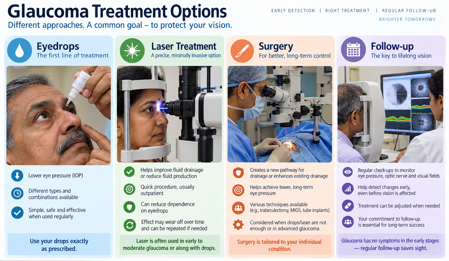
Glaucoma treatment aims to protect remaining vision. Most treatments work by lowering eye pressure, either by reducing fluid production or helping fluid drain better. The target pressure is different for each person. It depends on the type of glaucoma, starting pressure, optic nerve damage, age, and rate of progression.
Eye drop medicines
Eye drops are often the first treatment for open-angle glaucoma. They need regular use, usually for years. Missing doses can allow pressure to rise again.
Common medicine groups include:
Prostaglandin analogues
These usually help fluid drain better. They are often used once daily.
Beta blockers
These reduce fluid production in the eye. They may not suit people with certain breathing or heart conditions.
Carbonic anhydrase inhibitors
These lower fluid production and may come as drops or tablets in some cases.
Alpha agonists
These can reduce fluid production and improve drainage.
Combination drops
These combine two medicines in one bottle and may reduce the number of drops needed.
Eye drops can cause stinging, redness, dry eye, darkening of the iris or eyelid skin, eyelash changes, or effects elsewhere in the body. Patients should tell their doctor about asthma, heart disease, pregnancy, allergies, and all medicines they use.
A simple technique can reduce side effects. After putting in a drop, gently close the eye and press the inner corner near the nose for a minute. This can reduce the amount of medicine draining into the throat.
Laser treatment
Laser procedures can help in several situations.
Selective laser trabeculoplasty may be used for open-angle glaucoma. It treats the eye’s drainage tissue to improve fluid outflow. Some people use it to reduce dependence on drops, while others use it when drops do not control pressure well enough.
Laser peripheral iridotomy is often used for narrow angles or angle-closure glaucoma. It creates a tiny opening in the iris, helping fluid move and reducing the chance of sudden blockage.
Laser treatment is usually done without hospital admission, but follow-up remains needed. Its effect can reduce over time in some people.
Surgical options
Surgery is considered when medicines and laser do not control pressure enough, when the disease is advanced, or when a person cannot tolerate drops.
Common surgical approaches include:
Trabeculectomy
This creates a new drainage pathway for fluid to leave the eye.
Glaucoma drainage devices
These small implants help move fluid out of the eye to lower pressure.
Minimally invasive glaucoma surgery
These procedures use tiny devices or openings to improve drainage. They may suit selected patients, often with milder or moderate disease.
Cyclodestructive procedures
These reduce fluid production by treating the part of the eye that makes aqueous humour. They are often reserved for harder-to-control cases.
Every procedure has benefits and risks, including infection, bleeding, low pressure, cataract progression, scarring, or the need for more treatment. The right choice depends on the eye, the glaucoma type, and the level of pressure reduction required.

Daily habits and follow-up care
Lifestyle changes cannot replace glaucoma treatment, but routine habits can support eye health and treatment success.
Helpful steps include:
Use medicines exactly as prescribed
Keep follow-up appointments even when vision feels normal
Tell the doctor about side effects instead of stopping drops suddenly
Share a family history of glaucoma with relatives
Protect the eyes during sports, work, and home repairs
Manage diabetes, blood pressure, and other health conditions
Avoid unsupervised steroid use, especially around the eyes
People with glaucoma should also ask whether they are safe to drive, especially if visual field loss is present. Good central vision does not always mean side vision is adequate.
The key takeaway
Glaucoma is common, serious, and often quiet in its early stages. The main forms include primary open-angle glaucoma, primary angle-closure glaucoma, and secondary glaucoma caused by another eye condition, medicine, or injury. Risk rises with age, family history, raised eye pressure, steroid use, certain eye shapes, and some medical conditions.
A full diagnosis needs more than an eye pressure reading. The most useful assessments include optic nerve examination, visual field testing, OCT imaging, gonioscopy, and corneal thickness measurement.
Treatment can include eye drops, laser procedures, and surgery. The goal is to preserve the vision that remains. For many people, that is possible with early detection, regular monitoring, and consistent care. If glaucoma runs in the family or an eye check is overdue, booking a comprehensive eye examination is a sensible next step.



Comments